
 Anatomy set of muscled limbs : left leg and right arm
by
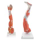
Part 1 Arm : This muscled arm model illustrates both the superficial and deeper muscles, five of which are removable from the muscled arm. Tendons, vessels, nerves and bone components of the left arm and shoulder are shown in great detail on this high quality muscle model. Parts numbered on muscled arm for easy identification of parts. - from distributors website.
Part 2 Leg: The muscular leg model illustrates both the superficial and deeper muscles, eight of which are removable. Tendons, vessels, nerves and bone components of the left leg and foot are shown in great detail in the muscular leg. All parts of muscular leg numbered. Muscular leg delivered on removable stand. -- from distributors website.
Anatomy set of muscled limbs : left leg and right arm
by
Part 1 Arm : This muscled arm model illustrates both the superficial and deeper muscles, five of which are removable from the muscled arm. Tendons, vessels, nerves and bone components of the left arm and shoulder are shown in great detail on this high quality muscle model. Parts numbered on muscled arm for easy identification of parts. - from distributors website.
Part 2 Leg: The muscular leg model illustrates both the superficial and deeper muscles, eight of which are removable. Tendons, vessels, nerves and bone components of the left leg and foot are shown in great detail in the muscular leg. All parts of muscular leg numbered. Muscular leg delivered on removable stand. -- from distributors website.
 Auditory ossicles
by
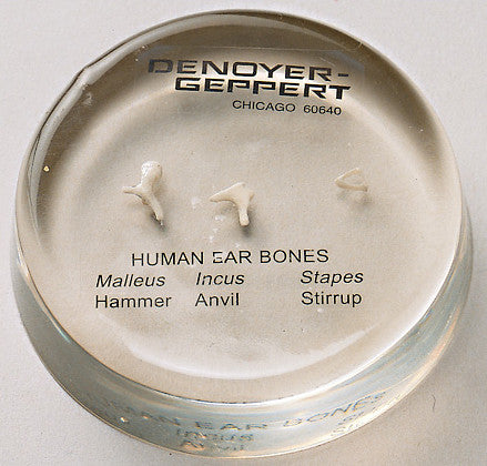
Life-Size replicas of the three tiny bones of the middle ear, the malleus (hammer), incus (anvil) and stapes (stirrup) cast from actual human specimens. Ossicles are labeled and embedded in plastic for protection.
Auditory ossicles
by
Life-Size replicas of the three tiny bones of the middle ear, the malleus (hammer), incus (anvil) and stapes (stirrup) cast from actual human specimens. Ossicles are labeled and embedded in plastic for protection.
 Axis Scientific 1.5 times life-size deluxe 4-part brain
by
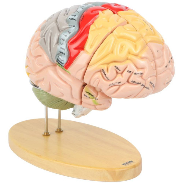
Anatomically accurate brain displayed at 1.5 times normal size, color coded with descriptive labels written on the model. Separates into 4 pieces, allowing for visibility to internal structures. Sits upon sturdy metal and wooden base.
Axis Scientific 1.5 times life-size deluxe 4-part brain
by
Anatomically accurate brain displayed at 1.5 times normal size, color coded with descriptive labels written on the model. Separates into 4 pieces, allowing for visibility to internal structures. Sits upon sturdy metal and wooden base.
 Axis Scientific Oversized 3-part human ear (5x life size)
by
With this oversized and detailed model of the human ear from Axis Scientific, it is easy to thoroughly study the internal structures of this crucial organ. The model is roughly 5 times life size, allowing for observation of even the tiniest structures of the ear.
Axis Scientific Oversized 3-part human ear (5x life size)
by
With this oversized and detailed model of the human ear from Axis Scientific, it is easy to thoroughly study the internal structures of this crucial organ. The model is roughly 5 times life size, allowing for observation of even the tiniest structures of the ear.
 Brain [model]
by
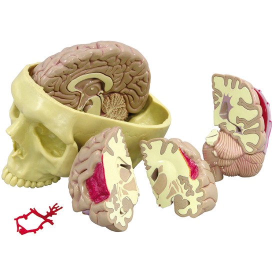
Model is a full size cut-away of the brain and shows the basic anatomy of the brain including illustrating how common pathologies might appear in the brain. The accompanying card contains the labelled display of what is shown by the model.
Brain [model]
by
Model is a full size cut-away of the brain and shows the basic anatomy of the brain including illustrating how common pathologies might appear in the brain. The accompanying card contains the labelled display of what is shown by the model.
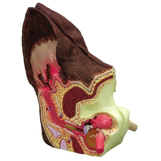 Canine ear model [model]
by
Model features normal anatomy and structures on one side and pathologies, such as inflammation, ear canal with partial occlusion and infection in bulla, on the other side.
Canine ear model [model]
by
Model features normal anatomy and structures on one side and pathologies, such as inflammation, ear canal with partial occlusion and infection in bulla, on the other side.
 Canine jaw [model]
by
Depicts healthy teeth on the right side and diseased/damaged teeth on the left side. Features nine pathologies: fractured canine, periodontal disease, tartar accumulation, plaque, gingivitis, worn incisors, retained deciduous tooth, gingival recession and missing premolar. The jaw may be opened, closed and separated for closer study.
Canine jaw [model]
by
Depicts healthy teeth on the right side and diseased/damaged teeth on the left side. Features nine pathologies: fractured canine, periodontal disease, tartar accumulation, plaque, gingivitis, worn incisors, retained deciduous tooth, gingival recession and missing premolar. The jaw may be opened, closed and separated for closer study.
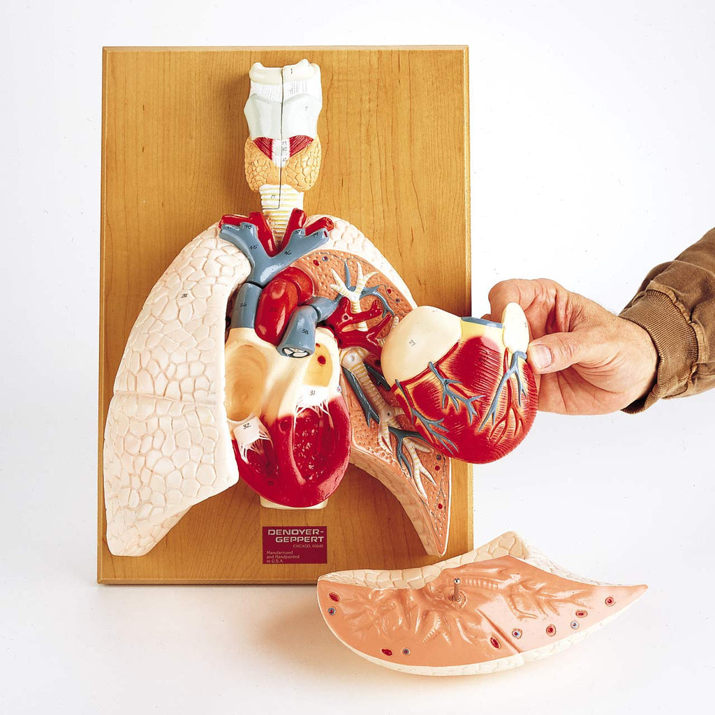 Cardiopulmonary system [model] : heart and respiratory organs
by
Shows lungs, trachea, heart, esophagus, and complete larynx with vocal cords in their natural positions. Exposes the bifurcation off the trachea and bronchial tree, and the pulmonary arteries and veins, when other organs are removed. Includes a 2-part heart that shows its four chambers and valves, thus providing understanding of the directional blood flow there. Allows tracing of pulmonary circulation and depicts major vessels of the systemic circulation to demonstrate blood flow through the entire body. Identifies 58 numbered structures in the guide.
Cardiopulmonary system [model] : heart and respiratory organs
by
Shows lungs, trachea, heart, esophagus, and complete larynx with vocal cords in their natural positions. Exposes the bifurcation off the trachea and bronchial tree, and the pulmonary arteries and veins, when other organs are removed. Includes a 2-part heart that shows its four chambers and valves, thus providing understanding of the directional blood flow there. Allows tracing of pulmonary circulation and depicts major vessels of the systemic circulation to demonstrate blood flow through the entire body. Identifies 58 numbered structures in the guide.